Business Wire10.24.17
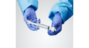
Stryker’s Spine division will introduce its Tritanium C Anterior Cervical Cage, a 3D-printed interbody fusion cage intended for use in the cervical spine, at the North American Spine Society (NASS) Annual Meeting, Oct. 25-28, 2017, in Orlando, Fla. (booth No. 500).
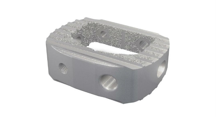
The Tritanium C Anterior Cervical cage is the newest addition to Stryker’s expanding line of spinal implants constructed from its proprietary Tritanium Technology,1 a novel, highly porous titanium material designed for bone in-growth and biological fixation.1 The unique porous structure of Tritanium is created to provide a favorable environment for cell attachment and proliferation, as demonstrated in an in-vitro study,2* and the Tritanium material may be able to wick or retain fluid, in comparison to traditional titanium.3 It is inspired by the microstructure of cancellous bone4 and enabled by AMagine, Stryker’s proprietary approach to implant creation using additive manufacturing, also known as 3D printing.
“After the terrific feedback and success we’ve seen with the Tritanium PL Posterior Lumbar cage, we are excited to introduce the Tritanium C Anterior Cervical Cage to spine surgeons this year at NASS,” said Bradley Paddock, President of Stryker’s Spine division. “Tritanium Cages feature ‘precisely randomized’4 pore formations, in contrast to other technologies with longitudinal channels and traverse windows that result in a uniform structure, as well as cages that offer porosity only on the surface. As a result, Tritanium implants are designed to become ‘one with bone’.”
“The ultimate goal with spinal implants is to get a solid fusion,” said Dr. Jocelyn Idema, a spine surgeon with Advanced Orthopaedics and Rehabilitation in Pittsburgh and Washington, Pa. “Spinal implants created with Tritanium Technology mimic the porosity of cancellous bone, which aids in fusion.”
Stryker’s Spine division also will present an abstract at NASS, titled, “Evaluation of Bony Fusion with Tritanium PL Used in Mini-Open Approach to Posterior Lumbar Interbody Fusion,” on October 26 at 1:41 p.m. ET, during the Innovative Technology Presentations.
The Tritanium C Anterior Cervical Cage received 510(K) clearance from the U.S. Food and Drug Administration in September 2017. It features an open central graft window and lateral windows to help reduce stiffness of the cage and minimize subsidence. In addition, the large graft window allows for bone graft containment. Engineered for stability,5,6 the cage has precisely angled teeth on the superior and inferior surfaces designed for bidirectional fixation and to maximize surface area for endplate contact with the cage. Its smooth posterior edges are designed to facilitate insertion and protect soft tissue and anatomy. The Tritanium C Anterior Cervical Cage is offered in a number of footprints, heights, and lordotic angles to accommodate a variety of patient anatomies.
The Tritanium C Cage is indicated for use in cervical interbody fusion procedures in skeletally mature patients with degenerative disc disease (DDD) at one level or two contiguous levels from the C2 to T1 disc. The cage is to be used with autogenous and/or allogenic bone graft comprised of cancellous and/or corticocancellous bone graft, and is to be implanted via an open, anterior approach. For the full Indications for Use, please refer to the Tritanium C Anterior Cervical Cage Instructions for Use.
References
1PROJ43909 Tritanium technology claim support memo
2RD0000053710: Tritanium cell infiltration and attachment experiment
*No correlation to human clinical outcomes has been demonstrated or established
3RD0000050927: Tritanium material capillary evaluation
4Karageorgiou V, Kaplan D. Porosity of 3D biomaterial scaffolds and osteogenesis. Biomaterials, 26, 5475-5491
5PROJ0000054458 | Tritanium C Insertion and Expulsion Marketing Memo.
6PROJ44960: Coefficient of friction memo