Michael Barbella, Managing Editor10.31.22
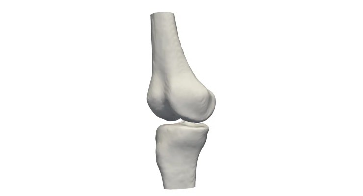
RSIP Vision is touting positive preliminary results from an ongoing clinical study on reconstructing a 3D knee model from 2D X-ray images. The tool is based on novel, patent-pending technology, implementing advanced artificial intelligence (AI) to produce an accurate knee model from two X-ray images (anteroposterior and lateral views). This model can be used for surgical planning and navigation during knee orthopedic procedures, such as total knee replacement.
“Robotic and patient-specific solutions for total knee arthroplasty (TKA) often require a CT scan for accurate 3D reconstruction of the knee bones.” said Moshe Safran, U.S. CEO at RSIP Vision. “Our knee reconstruction solution utilizes two standard X-ray images to produce a 3D model of the knee joint, comparable to the CT-based models. Our preliminary clinical study results demonstrate high accuracy, which we expect will be suitable for clinical applications. We believe our solution can alleviate the need for a knee CT-scan for TKA preoperative planning, providing access to precision planning for a much wider patient population.”
The goal behind developing this tool is to produce a high-grade, 3D knee model for surgical planning or navigation, with two main benefits; clinical and operational. The clinical benefit is lower levels of radiation exposure from X-ray imaging compared to a complete knee CT scan. Operationally, X-ray imaging has better accessibility than CT, is more widely reimbursed in the U.S. healthcare system, and is often lower in cost. To address the challenge of 3D reconstruction from standard X-ray images, both the unique AI technology and a custom tailored, low-cost calibration device developed by RSIP Vision are employed. After acquisition of AP and lateral X-rays, the software automatically creates a detailed 3D model of the knee joint, which can then be used for surgical planning.
The clinical evaluation of this tool is currently being conducted in Assuta Medical Center in Tel-Aviv, Israel. The system's inputs are two knee X-rays: anterior-posterior and lateral. The system automatically produces a 3D model of the femur and tibia. The tool's accuracy can be measured by comparing the resulting 3D models to the ground truth patient anatomy given in a corresponding CT scan. RSIP Vision plans to expand this clinical study into a U.S. medical center to ultimately receive U.S. Food and Drug Administration clearance. In parallel, RSIP Vision is working to extend this solution to additional anatomical regions such as the hip and shoulder joints.
“Patient specific surgical planning, especially in cases with bone deformity or anatomical variations, requires an accurate 3D model of the knee for implant assessment and procedural approach planning,” said Dr. Shai Factor, an orthopedic surgeon from Tel-Aviv Medical Center. “The CT scan is a good solution, but using X-ray images instead, is more feasible and will speed up the preoperative preparation process, while at the same time reducing radiation exposure. This new tool will provide the physician with a state-of-the-art 3D model of the knee without the need for a CT scan.”
RSIP Vision, led by Ron Soferman, develops computer vision and image processing R&D. In its 25 years of operation, RSIP Vision has provided clients with advanced customized software for their core businesses, using sophisticated algorithms and machine learning techniques. RSIP Vision employs engineers from various fields, including physics, computer science, mathematics, biomedicine, and neuroscience. Its engineers’ multidisciplinary expertise allows the firm to provide solutions.
