Julien Vidal, CEO of AZmed06.19.23
Designed to significantly improve the efficiency of radiologists, Deep Learning algorithm systems have become increasingly successful at detecting fractures, traumas, and abnormalities in X-Rays. The innovation comes at a time when medical imaging exams have doubled in OECD countries over the last 10 years but the number of specialists available to analyze the flow of information has stagnated. The technology is meant to automate many of the repetitive and time-consuming tasks physicians face in their day-to-day so more time is spent caring for patients and constructing a personalized treatment plan.
In a 2019 study performed in Louis Mourier Emergency Room in France, out of 25 patients diagnosed by the clinician as having a fracture, the algorithm was able to detect fractures in 24 of those patients.
An algorithm designed to act similarly to how the brain functions, Deep Learning, a subfield of Artificial Intelligence, is able to analyze data without supervision. Since 2012, it has enhanced the medical imaging analysis process and has decreased the classification error rate from about 25% in 2011 to 3.6% in 2015. The technology has been applied in a number of fields like detecting skin cancer, mammograms, and diabetic retinopathy.
Across the 21 centers part of University Hospitals Cleveland, 2,626 radiographs were analyzed along with 16 radiologists and eight emergency room physicians. On average, doctors using the DL algorithm system saved 27% less time reviewing imaging.
While not designed to eliminate the need for a medical professional completely, innovations like these are designed as a safety net for a more secure diagnosis after a significant increase in the number of imaging examinations since the 1990s. Looking into the next few years, AI detectors will be able to assist physicians in diagnosing more than just X-rays and fractures as Rayvolve is already able to accurately locate traumas in bones as well as pulmonary and cardiac pathologies.
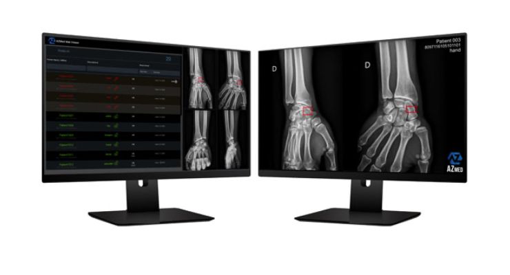
In designing this type of software, easy accessibility and seamless usability is a necessity. Rayvolve is integrated into the existing radiology Picture Archiving and Communication System (PACS) reading workflow. Predictions are generated automatically in the same series of Digital Imaging and Communications in Medicine (DICOM) exams.
At the same time, as AI aids are becoming increasingly more intelligent, imaging exams are able to see more of the human body than ever before. For the more common ailments, the coming wave of AI tools will significantly streamline diagnoses and help patients get quality care quicker. This will also allow for trained professionals to have dedicated time to think through more challenging imagining analyses, reduce the risk of misdiagnoses and offer more in-depth face-to-face time with patients.
Co-founder of AZmed, Julien Vidal along with his team aim to optimize physicians’ workflow and augment workflows through AI in order to ensure doctors spend enough time on what matters most: patients. Julien along with his co-founders were all nominated for Forbes 2022 “30 Under 30” and AZmed is the first French company to obtain the CE mark for Artificial Intelligence software in conventional radiology. The software is FDA-cleared and is now used in more than 33 countries, 900 healthcare institutions and has been adopted by industry leaders such as the NHS and Amazon Web Services. FII Institute, a non-profit organization committed to ESG principles, invested in the French start-up in 2021 which stands as a testament to its mission of catalyzing projects to move beyond research and dialogue to secure real-world solutions.
In a 2019 study performed in Louis Mourier Emergency Room in France, out of 25 patients diagnosed by the clinician as having a fracture, the algorithm was able to detect fractures in 24 of those patients.
An algorithm designed to act similarly to how the brain functions, Deep Learning, a subfield of Artificial Intelligence, is able to analyze data without supervision. Since 2012, it has enhanced the medical imaging analysis process and has decreased the classification error rate from about 25% in 2011 to 3.6% in 2015. The technology has been applied in a number of fields like detecting skin cancer, mammograms, and diabetic retinopathy.
Industry Application
A natural next step in developing this innovation is having a seamless process for physicians to access the system without having to change their working habits. AZmed’s Rayvolve system has a 99% sensitivity rate, meaning the system has a very strong likelihood of detecting abnormalities and an 89% specificity rate. It has also avoided false negatives by 67%. As with most Deep Learning algorithms, as the system is exposed to more data over time, the more accurate and efficient the technology becomes at identifying fractures and abnormalities in X-rays. Rayvolve is the first software of its kind to obtain the CE Mark in its category which indicates that the product has been assessed by the manufacturer and deemed to meet EU safety, health and environmental protection requirements.Across the 21 centers part of University Hospitals Cleveland, 2,626 radiographs were analyzed along with 16 radiologists and eight emergency room physicians. On average, doctors using the DL algorithm system saved 27% less time reviewing imaging.
While not designed to eliminate the need for a medical professional completely, innovations like these are designed as a safety net for a more secure diagnosis after a significant increase in the number of imaging examinations since the 1990s. Looking into the next few years, AI detectors will be able to assist physicians in diagnosing more than just X-rays and fractures as Rayvolve is already able to accurately locate traumas in bones as well as pulmonary and cardiac pathologies.
In designing this type of software, easy accessibility and seamless usability is a necessity. Rayvolve is integrated into the existing radiology Picture Archiving and Communication System (PACS) reading workflow. Predictions are generated automatically in the same series of Digital Imaging and Communications in Medicine (DICOM) exams.
At the same time, as AI aids are becoming increasingly more intelligent, imaging exams are able to see more of the human body than ever before. For the more common ailments, the coming wave of AI tools will significantly streamline diagnoses and help patients get quality care quicker. This will also allow for trained professionals to have dedicated time to think through more challenging imagining analyses, reduce the risk of misdiagnoses and offer more in-depth face-to-face time with patients.
Co-founder of AZmed, Julien Vidal along with his team aim to optimize physicians’ workflow and augment workflows through AI in order to ensure doctors spend enough time on what matters most: patients. Julien along with his co-founders were all nominated for Forbes 2022 “30 Under 30” and AZmed is the first French company to obtain the CE mark for Artificial Intelligence software in conventional radiology. The software is FDA-cleared and is now used in more than 33 countries, 900 healthcare institutions and has been adopted by industry leaders such as the NHS and Amazon Web Services. FII Institute, a non-profit organization committed to ESG principles, invested in the French start-up in 2021 which stands as a testament to its mission of catalyzing projects to move beyond research and dialogue to secure real-world solutions.
