Pratik Kirve, Sr. Specialist—Content Writer, Allied Analytics LLP08.07.19
Healthcare professionals have always been looking for advanced technologies that can offer critical information during surgeries and enable efficiency and better results. There is a huge demand for minimally invasive procedures, and advanced technologies would facilitate those procedures. Technologically advanced surgical imaging systems have been introduced and implemented in healthcare facilities to gain various benefits. New systems reduce exposure to radiation and improve results of surgeries. Mercy Medical Center recently implemented a multi-dimensional imaging platform that can be used in spine, cranial, and trauma-related surgeries. It can also be used for minimally invasive spinal procedures. These systems offer faster and better results as compared to other conventional systems.
New advanced systems offer the crucial feature of real-time information. This enables surgeons to take necessary steps immediately and offer better care. This also saves patient costs and enables optimum outcomes. Healthcare facilities have been rapidly adopting the advanced systems—the demand for advanced imaging systems is immense, leading to a rise in the market. According to a report by research firm Allied Market Research, the global surgical imaging market is expected to reach $1.49 billion by 2026. The implementation of new technologies in imaging systems have yielded better results in terms of patient safety and efficient procedures.
Improving surgery results through imaging systems equipped with advanced technology is crucial for medical centers. Mercy Medical Center recently installed the Medtronic O-arm Mobile Surgical Imaging System to reduce exposure to radiation and enhance spinal surgery results. The imaging system is a multi-dimensional imaging platform used in spine, cranial, and trauma-related surgeries. It offers intraoperative and real-time imaging of bones and implants with high quality images. Moreover, it provides a large field-of-view in two and three dimensions. Charles C. Park, M.D., Ph.D., Director, The Minimally Invasive Brain and Spine Center, Mercy Medical Center, highlighted that the O-arm, a mobile X-ray machine, provides a 360-degree scan of the patient’s anatomy during spinal, thoracic, lumbar, and cervical surgeries. Along with offering excellent viewing positions, it enables real-time navigation of spinal instrumentation in 3D. Though the O-arm is utilized in traditional and open surgeries, Dr. Park uses it for minimally invasive spinal procedures. He added the system offers precision in hardware placement. Moreover, it scans the entire cervical spine in only 13 seconds. It has an ability to capture nearly 400 images in less than 30 seconds. The results are fast, and the system enables less invasive surgery in less time.
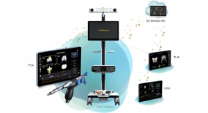
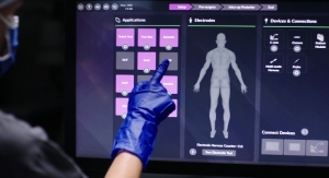
Similarly, NuVasive's Pulse OR platform offers a combination of surgical planning, neuromonitoring, radiation reduction, rod bending, and imaging and navigation functions to enhance operational workflow, lessen variability, and raise surgical reproducibility. The independent access enables image viewing from various technologies and provides insights in real time. An open and modular architecture of the system enables usage of flexible technologies.
The Pulse OR platform’s hardware and software modules offer image transfer for both 2D- and 3D-imaging and joins camera, array, and workflow technologies for better line of sight, ease-of-use, and surgical efficiency. It also offers reduction in radiation through LessRay, an imaging system that can take low-dose, low-quality images and improve them to make them look like full-dose images. Juan Uribe, M.D., chief of the division of spinal disorders at Barrow Neurological Institute, commented that NuVasive’s Pulse platform offers an incredible surgical experience with its optimized and responsive workflow features. It provides the benefits of the advanced imaging and navigation along with neuromonitoring that enables optimized outcomes for patients.
The advent of new technologies has helped hospitals improve patient safety and efficiently carry out surgeries. All India Institute of Medical Science (AIIMS) in Delhi, India installed the first fluorescence imaging technology for ease in identification of cancer-affected tissues in breasts. Dr. SVS Deo, Head of the Department of Surgical Oncology at AIIMS, outlined that this technology would prove to be revolutionary for breast cancer surgery because it offers precision in determining cancer-affected tissues intraoperatively. A safe and affordable indocyanine green (ICG) dye is injected in patients during the breast cancer surgery. With the help of fluorescence imaging technology, surgeons can see blood flow in micro-vessels, vessels, tissue perfusion, and anatomical structures in an intraoperative manner. The affected tissues show a fluorescent green color. The new technology differs from other conventional technologies in both reliability and offering multiple imaging applications.
Dr. Deo added all the lymph nodes—including healthy ones—were removed due to unavailability of critical information, which caused collateral damage to patients. Fluorescence imaging technology was able to help save healthy tissues and improve patient safety. The near-infrared fluorescence imaging technology was used to enable real-time, actionable information, improving patients’ quality of care and safety. Dr. David Weintritt, a breast cancer expert from GW School of Medicine and Health Sciences, backed Dr. Deo’s comments. He said this technology offered critical patient anatomical information when the timing was most crucial—adding that several complications can be avoided and the healthcare burden can be reduced.
Dr. Weintritt also explained the technology can be utilized in breast oncoplasty along with breast reconstruction post-mastectomy. It also shows the areas that do not contain enough blood supply, so the surgeon can eliminate tissues that may cause infections or healing problems and lead to unnecessary additional expensive surgeries.
Pratik Kirve is a writer, blogger, and sports enthusiast. He holds a bachelor degree in Electronics and Telecommunication Engineering and currently works as a Sr. Specialist - Content Writer at Allied Analytics LLP. He has avid interest in writing news articles across different verticals.
New advanced systems offer the crucial feature of real-time information. This enables surgeons to take necessary steps immediately and offer better care. This also saves patient costs and enables optimum outcomes. Healthcare facilities have been rapidly adopting the advanced systems—the demand for advanced imaging systems is immense, leading to a rise in the market. According to a report by research firm Allied Market Research, the global surgical imaging market is expected to reach $1.49 billion by 2026. The implementation of new technologies in imaging systems have yielded better results in terms of patient safety and efficient procedures.
Improving surgery results through imaging systems equipped with advanced technology is crucial for medical centers. Mercy Medical Center recently installed the Medtronic O-arm Mobile Surgical Imaging System to reduce exposure to radiation and enhance spinal surgery results. The imaging system is a multi-dimensional imaging platform used in spine, cranial, and trauma-related surgeries. It offers intraoperative and real-time imaging of bones and implants with high quality images. Moreover, it provides a large field-of-view in two and three dimensions. Charles C. Park, M.D., Ph.D., Director, The Minimally Invasive Brain and Spine Center, Mercy Medical Center, highlighted that the O-arm, a mobile X-ray machine, provides a 360-degree scan of the patient’s anatomy during spinal, thoracic, lumbar, and cervical surgeries. Along with offering excellent viewing positions, it enables real-time navigation of spinal instrumentation in 3D. Though the O-arm is utilized in traditional and open surgeries, Dr. Park uses it for minimally invasive spinal procedures. He added the system offers precision in hardware placement. Moreover, it scans the entire cervical spine in only 13 seconds. It has an ability to capture nearly 400 images in less than 30 seconds. The results are fast, and the system enables less invasive surgery in less time.
Similarly, NuVasive's Pulse OR platform offers a combination of surgical planning, neuromonitoring, radiation reduction, rod bending, and imaging and navigation functions to enhance operational workflow, lessen variability, and raise surgical reproducibility. The independent access enables image viewing from various technologies and provides insights in real time. An open and modular architecture of the system enables usage of flexible technologies.
The Pulse OR platform’s hardware and software modules offer image transfer for both 2D- and 3D-imaging and joins camera, array, and workflow technologies for better line of sight, ease-of-use, and surgical efficiency. It also offers reduction in radiation through LessRay, an imaging system that can take low-dose, low-quality images and improve them to make them look like full-dose images. Juan Uribe, M.D., chief of the division of spinal disorders at Barrow Neurological Institute, commented that NuVasive’s Pulse platform offers an incredible surgical experience with its optimized and responsive workflow features. It provides the benefits of the advanced imaging and navigation along with neuromonitoring that enables optimized outcomes for patients.
The advent of new technologies has helped hospitals improve patient safety and efficiently carry out surgeries. All India Institute of Medical Science (AIIMS) in Delhi, India installed the first fluorescence imaging technology for ease in identification of cancer-affected tissues in breasts. Dr. SVS Deo, Head of the Department of Surgical Oncology at AIIMS, outlined that this technology would prove to be revolutionary for breast cancer surgery because it offers precision in determining cancer-affected tissues intraoperatively. A safe and affordable indocyanine green (ICG) dye is injected in patients during the breast cancer surgery. With the help of fluorescence imaging technology, surgeons can see blood flow in micro-vessels, vessels, tissue perfusion, and anatomical structures in an intraoperative manner. The affected tissues show a fluorescent green color. The new technology differs from other conventional technologies in both reliability and offering multiple imaging applications.
Dr. Deo added all the lymph nodes—including healthy ones—were removed due to unavailability of critical information, which caused collateral damage to patients. Fluorescence imaging technology was able to help save healthy tissues and improve patient safety. The near-infrared fluorescence imaging technology was used to enable real-time, actionable information, improving patients’ quality of care and safety. Dr. David Weintritt, a breast cancer expert from GW School of Medicine and Health Sciences, backed Dr. Deo’s comments. He said this technology offered critical patient anatomical information when the timing was most crucial—adding that several complications can be avoided and the healthcare burden can be reduced.
Dr. Weintritt also explained the technology can be utilized in breast oncoplasty along with breast reconstruction post-mastectomy. It also shows the areas that do not contain enough blood supply, so the surgeon can eliminate tissues that may cause infections or healing problems and lead to unnecessary additional expensive surgeries.
Pratik Kirve is a writer, blogger, and sports enthusiast. He holds a bachelor degree in Electronics and Telecommunication Engineering and currently works as a Sr. Specialist - Content Writer at Allied Analytics LLP. He has avid interest in writing news articles across different verticals.