Michael Barbella, Managing Editor02.12.18
Athleticism runs deep in the Strachan family bloodline.
Three generations of the Waterbury, Conn.-based clan have produced both football and cheerleading coaches as well as an all-American high school cheerleader. It also bore a trauma nurse—a byproduct, perhaps, of the unspoken brutality and dangers associated with contact sports.
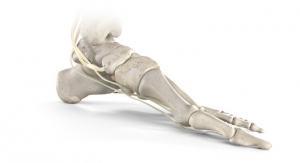
That lineage, however, was nearly exterminated two years ago by a devastating injury to the Strachan dynasty’s only remaining heir. Leah Strachan, (then) 15, was in the midst of a routine cheer practice when she landed on the mat to crushing pain in her left ankle.
Strachan was convinced she’d broken a bone: Her ankle ballooned beyond recognition, grew increasingly stiff, and most tellingly, couldn’t support her weight. She feared her cheerleading days were over.
But X-rays ruled out a fracture, leaving Strachan, her family and her puzzled doctors with more questions than answers. Strachan’s search for a solution (more of a diagnosis, really) beget numerous clinical consultations, analytical tests, and frustrating dead-ends, though it did ultimately yield the specialist who eventually would orchestrate her odds-defying recovery.
Raymond J. Walls, M.D., a Yale School of Medicine surgeon and assistant professor of orthopedics and rehabilitation, diagnosed Strachan’s injury as a combination ankle osteochondral lesion (joint surface damage) and ligament rupture. Adding to the misery was chronic inflammation, scar tissue buildup, and a loose bone fragment inside the ankle.
It was, strictly speaking, quite the messy injury.
After exhausting all nonsurgical solutions, Walls decided to rebuild Strachan’s damaged ankle naturally, using the teenager’s own stem cells to promulgate healing. “It’s a bit like adding nitro to a car engine,” Walls said in a Yale Medicine website feature story on Strachan’s recovery. “We use the patient’s own reparative body cells in a high concentration to greatly enhance the natural healing process.”
Harnessing Mother Nature’s magic has progressively gained traction among orthopedic surgeons in recent years as they seek better treatment options for injured cartilage, ligaments, tendons, and joints. Their efforts have given rise to a lucrative biologics market, estimated to grow 5.4 percent annually over the next four years to reach $6.06 billion in 2022 from $4.66 billion last year, according to Research and Markets data.
Scientists have long been aware of the human skeleton’s remarkable regenerative abilities, but have traditionally fallen short in their attempts to manipulate physics. They have found considerable success, however, with remedies like platelet-rich plasma (PRP) and bone marrow aspirate concentrate (BMAC)—therapies that use a patient’s own blood and other tissues to repair defective joints.
First used by oral surgeons to regenerate bone and soft tissue in the jaw, PRP is a blood-based biologic that contains a high concentration of protein-rich platelets. Studies suggest the growth factors released by platelets recruit reparative cells, augment tissue repair, and help accelerate soft tissue healing. PRP has benefited busted rotator cuffs and Achilles tendons as well as chronically injured tendons that refuse to heal properly.
PRP is not sanctioned by the U.S. Food and Drug Administration (FDA), and its long-term efficacy has yet to be definitively proven. Nevertheless, famous athletes like Tiger Woods and Rafael Nadal have used PRP injections to help heal their injuries.
“It’s [about] repair and regeneration rather than removal and replacement,” orthopedic surgeon Martha M. Murray, head of the Sports Medicine Research Laboratory at Boston Children’s Hospital, told the online web portal Health WorldNet last fall.
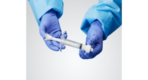
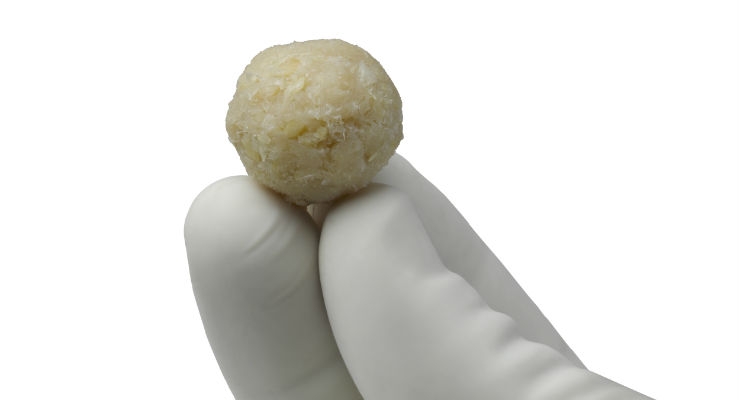
BMAC targets repair and regeneration through “undifferentiated” stem cells that can replicate themselves into various tissue types. The stem cells are extracted from the patient’s own bone marrow (via hip injection) and concentrated using a centrifuge to produce a dense clot-like “spackle” for filling gaps in cartilage and surrounding grafted tissue.
Walls used this technique to patch up Strachan’s ravaged ankle. He first removed the joint’s inflamed, thickened scar tissue, and extracted areas of ruined cartilage surface, which the Ireland-educated doctor likened to a “severely damaged portion of a rubber tire covering a car wheel.”
Next, Walls made small holes in the underlying bone surface to stimulate bleeding and fill the cartilage defect. Then, to maximize Strachan’s chances of a complete recovery, Walls combined the teen’s stem cells with a cartilage scaffold—an amalgamation shown in preclinical testing to enhance the formation of normal cartilage after injury. “We believe this will extend the lifespan of the repair tissue way beyond five years and hopefully last a lifetime,” Walls said.
While the longevity of Walls’ repair work won’t truly be determined for decades, initial results are promising: Strachan regained stability and almost the full range of motion in her ankle just six weeks after the surgery. Three months post-op, she participated in sports-specific rehabilitation (mostly low-impact exercises) and six months after surgery, she was back to performing gymnastics maneuvers. Now a high school senior, Strachan is “fully back” and eager to join a college cheerleading squad.
“...there are evolving uses of autologous platelet-rich plasma and bone marrow aspiration concentration in soft tissue and joint health applications,” noted Curtis Matthews, chief operating officer at Reinvent Biologics, a regenerative medicine developer and manufacturer based in Fort Worth, Texas. “Although these uses are not indicated, based on the 510(k) indication statements of the commercial products, physicians are using their own medical discretion. There are some exciting results that have been reported in increasingly more peer-reviewed journals.”
And those results transcend body parts. A pair of studies published in the American Journal of Sports Medicine in 2016, for instance, tout BMAC’s advantages for knee repair. In one trial, BMAC treatment (via hyaluronic acid scaffold) for grade IV chondral lesions produced better outcomes and more durable cartilage repair at medium-term follow-up (two and five years) than microfracture.
BMAC also has proven its worth in relieving pain and improving activity levels in patients battling both osteoarthritis and chronic patellar tendinopathy (“jumper’s knee”).
“With the goal being the development of products to aid in the effective and efficient healing of patients,” Matthews said, “advances are being made in differing processing techniques of donated human allografts, inductive properties of synthetics, and the concentration techniques of platelet-rich plasma and bone marrow concentration.”
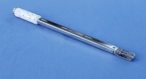
Progress in the latter category has spawned faster, more convenient centrifugation systems and higher cell concentration factors. The automated CellPoint system from St. Louis, Mo.-based Isto Biologics, for example, requires no operator intervention and achieves typical total nucleated cells (TNC) concentration factors that are three to six times greater than baseline bone marrow aspirate TNC concentration levels.
Harvest Technologies (a Terumo BCT company), meanwhile, claims its decade-old BMAC system beats five other competitors in stem cell concentration levels and yield. Moreover, the system reportedly simplifies training among multiple users and can produce autologous biologics in less than 15 minutes.
Reinvent Biologics’ Genius Concentration System reduces that processing time to two minutes and achieves an 84 percent retention of nuclear cells and an 86 percent retention of platelets when processing peripheral blood and bone marrow aspirate. This is achieved with a 3.2 percent coefficient of variance. The company launched the Genius system shortly after its founding in 2016 and expanded the product line last fall to include a larger-capacity option for blood and bone marrow aspirate processing.
In addition to the larger-capacity Genius system, Reinvent debuted two new product lines in 2017—the Einstein family of allograft bone (Inductive Bone Matrix, Demineralized Bone Matrix, and Compressive Bone Scaffold), and the Wizard line of synthetic bone graft substitutes comprised of beta-tricalcium phosphate (TCP) granules and a calcium phosphate flowable cement.
“Over the past few years, innovation in orthobiologics has been driven by the desire to discover a solution that is either equivalent or superior to the ‘gold standard’ in bone grafting, the iliac crest wedge harvest,” Matthews explained. “As co-morbidities such as increased harvest site pain, increased infection rates, and/or slower healing due to second site injury may exist when harvesting a wedge of iliac crest, researchers have been looking for an equivalent or superior solution without the significant risk of certain co-morbidities. The cycle of possible solutions has oscillated from synthetic bone graft extenders to pharmaceutical solutions, to differently processed human allografts, to different harvesting methods and processing of autologous grafts.”
That resolution cycle likely will persist well into the future, too, as science continues its quest to replicate the body’s natural bone-renewing ability. The crusade has generated a deluge of substitute bone graft material options from both OEMs and smaller firms, as well as an assortment of promising new breakthroughs from university researchers.
Clearly, the most controversial and well-known bone graft surrogate is Medtronic plc’s Infuse, a biologic agent approved by the FDA in 2002 for certain spine, oral-maxillofacial, and orthopedic trauma surgeries. Powered by a genetically engineered human bone growth protein, the product helps form new bone and has been used in more than 1 million patients over the last 15 1/2 years.
Clinical studies have shown a healing equivalency between Infuse and autografts in lumbar fusion surgery, but extensive off-label use of the product has triggered thousands of lawsuits and a U.S. Senate committee inquiry over safety concerns. Medtronic has paid millions of dollars to settle the lawsuits, and late last year, shelled out $12 million to resolve investigations in five states over its marketing strategy for Infuse.
A significantly less contentious alternative is offered by NovaBone Products LLC, developer of the industry’s first bioactive synthetic bone graft. Based on 30 years of research in bone regeneration, the firm’s self-named technology (NovaBone) is rooted in a calcium phosophosilicate composite that boosts the body’s ability to signal, recruit, proliferate, and differentiate bone-building cells—a process deemed by the FDA as “osteostimulation.”
“Orthobiologics has been evolving towards products that stimulate bone growth,” NovaBone President and CEO Arthur Wotiz told Orthopedic Design & Technology. “NovaBone products stimulate the body’s natural healing process rather than change it. Therefore, there are no issues with mimicking the body’s natural healing process for bone.”
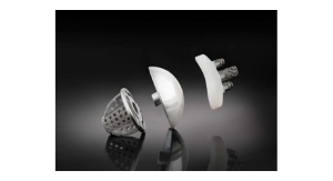
NovaBone Products’ bone graft substitutes come in various forms, including putty, morsels, particulate, a collagen bioactive glass scaffold, and a moldable, continuous porous structure, among others. Like its OEM rival, NovaBone’s technology has been used for more than a decade to repair osseous skeletal defects in over 1 million clinical applications.
Competing with NovaBone for orthobiologics pioneer bragging rights is Stryker Corp., which boasts more than 20 years of clinical research in regenerative technologies for bone, cartilage, and soft tissue injuries/defects; and DePuy Synthes, whose entry into the cellular allograft sector dates back nearly two decades.
Stryker’s biologics portfolio consists of Imbibe, a single-use, disposable harvesting system for bone marrow; Cortoss, an injectable, non-resorbable polymer composite designed to mimic cortical bone; HydroSet, an injectable, sculptable, self-setting bone substitute,; and Vitoss, a line of (purportedly) top-selling bone void fillers used in more than 425,000 implantations globally. The company also markets several soft tissue products, including hemostats and a bovine-derived type 1 collagen and intact dermis tissue.
DePuy’s stand-ins for autologous grafts include bio-compatible calcium phosphate bone void fillers in either fast-set putty form (Norian SRS) or strips (chronOS).
“The body is often able to heal itself with little or no help from biomaterials. In the case of very straightforward surgeries in healthy patients in which the native bone fragments are still intact, surgeons can use very simple bone void fillers such as chronOS Bone Graft Substitute or in some cases none at all,” said Matthew D. Putnam, M.D., franchise medical director at DePuy Synthes Companies. “The main objective of these types of surgeries is to have the bony anatomy restored to its pre-injury or desired alignment, fill in any small voids or gaps in the musculoskeletal system with a biomaterial, and then lock that down with hardware—from there the body does the rest.”
Indeed, activating the natural healing process is a cornerstone of orthobiologic treatment technology. But the methods employed to attain that goal are as varied as the companies competing in the space.
Westminster, Colo.-based Cerepedics Inc., for example, uses a synthetically derived 15-amino acid peptide (P-15) to support bone growth, while Scottish startup SIRAKOSS Ltd. leverages its proprietary expertise in granule chemistry and architecture to achieve the same outcome. Artoss GmbH, on the other hand, relies on nano-crystalline hydroxyapatite (HA) particles to jumpstart bone growth.
Zimmer Biomet’s bone stimulating formula also includes HA particles but mixes them with calcium sulphate to create a material (CERAMENT) that mimics the stiffness and strength of cancellous bone. The resulting calcium sulphate-HA matrix, according to a product data sheet, creates an osteoconductive framework that fosters new bone formation.
Non-HA solutions include an FDA-approved ß-tricalcium phosphate-porcine collagen brew from German biomaterials developer curasan AG and a calcium phosphate blend from French bioactive biomaterials manufacturer Graftys S.A. The latter innovation, cleared for use both in Europe and the United States, facilitates healthy bone reconstruction through an osteoconductive, porous, and permeable scaffold.
Graftys also is developing a clot-enhancing osteogenic putty comprised of autologous whole blood and ceramic. Initially shown to rival bone autograft in animal models, the material contains BCP (biphasic calcium phosphate) microparticles enriched with calibrated calcium ions for optimum osteo-conduction.
“The overall orthobiologics market continues to be fueled by the need for products that deliver enhanced, consistent, and predictable healing across various clinical indications,” said David Yonce, vice president of Innovation and R&D at DSM Biomedical, a global medical device materials developer and manufacturer. “Increased focus on demonstrated effectiveness and the recent clarification and guidance from the FDA regarding human cellular and tissue products, combined with the general declining use of allografts and autografts, is driving increased attention towards alternative synthetic technologies.”
DSM’s contribution to the world of biologic alternatives encompasses biabsorbable polymers (spine plating), ultra-high molecular weight polyethylene cerclage cables, extracellular matrices, and bioceramic-collagen bone grafts. Its signature product is an ultra-high molecular weight polyethylene fiber ideal for use in intricate and minimally invasive surgeries. Billed as the “world’s strongest fiber,” Dynema Purity fiber is 15 times stronger than steel, 40 percent tougher in weight than aramids, and three times heartier in volume than polyester.
Last spring, DSM released an ultra-high molecular weight polyethylene black fiber for use in various medical devices like surgical sutures and different-colored cables. The black fibers are particularly ideal for sports medicine applications, as they enable more complex sutures to be manufactured and help reduce the breakage commonly associated with polyester stitches.
“The next generation of orthobiologic technologies in development are based upon scientific and clinical knowledge gained through experience,” explained Yonce. “These insights provide better understanding of the tissue’s response from cell-signaling, material-biology interactions, and the implants’ surrounding environment. Accounting for these multi-factorial elements will contribute towards an improved physiologic balance and better healing conditions. We believe the next generation of orthobiologic materials will strike the appropriate balance of handling and structural properties while achieving the physiologic balance for accelerated healing.”
Many of those descendant materials will likely be born from academic research, too. One is already gestating at the University of Bristol in the United Kingdom: Researchers there are exploring whether a small lipid molecule called FHBP can accelerate the maturation of immature bone-forming stromal cells into osteoblasts on bone graft surfaces. The analysts tested the FHBP-modified grafts in human and sheep cell cultures spiked with active vitamin D3 and compared their performance with that of unmodified grafts.
Study data indicated the FHBP-coated grafts expedited bone-forming cell maturation on the graft surface but difference in responses between sheep and human cell cultures made the former an unsuitable animal model without further investigations.
More promising results are emerging from Northwestern University (Evanston, Ill.), where scientists are studying the in vivo effects of a “sugar-coated” nanomaterial on the activity of bone morphogenetic protein 2 (BMP-2). The nanomaterial consists of miniscule filaments that molecularly bind BMP-2 similar to the way natural sugars are bound in the body. The coated molecule is a synthesized sugar polymer known as sulfated polysaccharide, which resembles sugars used by nature to activate BMP-2 when cell signaling indicates bone growth.
The sugar molecules on the nanostructure’s surface “grab” the protein in the exact spot used in biological systems to deploy the bone-growing signal. The process, according to Northwestern researchers, increases bone-growing signals beyond the levels initiated by the body’s naturally occurring sugar polymers. University scientists are planning to seek FDA approval to launch a human clinical trial studying the nanomaterial for bone regeneration.
“We focused on bone regeneration to demonstrate the power of the sugar nanostructure to provide a big signaling boost,” said Samuel I. Stupp, who developed the new nanomaterial. “With small design changes, the method could be used with other growth factors for the regeneration of all kinds of tissues. One day we may be able to fully do away with the use of growth factors made by recombinant biotechnology and instead power the natural ones in our bodies.”
New Jersey Institute of Technology (NJIT) engineers have the same hope. A team at the school’s Tissue Engineering and Biomaterials Laboratory is developing a bioactive composite matrix designed to serve as a bone graft substitute. Resembling thick fabric, the matrix is a deformable, bioceramic fibrous material that can be cut with a pair of scissors for easy insertion into bone defects. The material has a high fiber structure with a high surface area to allow for cell stretching and anchoring. It also is highly porous to enable bone tissue growth.
NJIT’s bone matrix technology was originally developed through funding from the National Science Foundation and the Coulter Foundation, which funds translational studies. It also has received $200,000 in funding from both NJIT and the University City Science Center in Philadelphia, Pa.
Besides the bone matrix material, NJIT’s laboratory has pioneered the use of bioactive ceramics and composites for use in bone-tissue engineering. Bio-inspired substances such as glycosaminoglycan (GAG) mimetics and piezoelectric materials are currently being developed as well for bone, cartilage, and neurological applications. GAG mimetics combine with growth factors to simulate tissue growth and piezoelectric materials provide electrical stimulation to cells.
“It is challenging to mimic the body’s healing process for bone, as its function in providing support for the musculoskeletal system has to be balanced with the biology of bone regeneration,” noted Michael Carter, general manager of Stryker’s Spine Division. “Bringing this balance of biomechanics and biology to life requires advanced manufacturing techniques that were previously unavailable. Recent advancements in additive manufacturing, also known as 3D printing, have pushed beyond those limits and are driving innovation in previously unmanufacturable shapes that are designed to mimic the porosity of bone. For example, Stryker Spine’s portfolio of interbody cages is built with Tritanium technology, which is designed to mimic cancellous bone and was designed for bone in-growth and biological fixation.”
Hossein Montazerian is aiming to mimic Mother Nature through 3D printing as well. The University of British Columbia-Okanagan research assistant has identified a way to model and create custom-printed artificial bone grafts.
In his research, Montazerian identified 240 different bone graft designs but focused only on the ones that were both porous and strong. He used 3D printing to create the designs that performed best and then ran physical tests to determine their real-world efficacy. Results showed the best designs were up to 10 times stronger than the others and significantly less likely to cause long-term problems.
“We hope to produce bone grafts that will be ultra porous, where the bone and connective tissues meet and are extra strong at the points under the most stress,” he affirmed last summer. “The ultimate goal is to produce a replacement that almost perfectly mimics real bone.
A true challenge for the ages.
Three generations of the Waterbury, Conn.-based clan have produced both football and cheerleading coaches as well as an all-American high school cheerleader. It also bore a trauma nurse—a byproduct, perhaps, of the unspoken brutality and dangers associated with contact sports.
That lineage, however, was nearly exterminated two years ago by a devastating injury to the Strachan dynasty’s only remaining heir. Leah Strachan, (then) 15, was in the midst of a routine cheer practice when she landed on the mat to crushing pain in her left ankle.
Strachan was convinced she’d broken a bone: Her ankle ballooned beyond recognition, grew increasingly stiff, and most tellingly, couldn’t support her weight. She feared her cheerleading days were over.
But X-rays ruled out a fracture, leaving Strachan, her family and her puzzled doctors with more questions than answers. Strachan’s search for a solution (more of a diagnosis, really) beget numerous clinical consultations, analytical tests, and frustrating dead-ends, though it did ultimately yield the specialist who eventually would orchestrate her odds-defying recovery.
Raymond J. Walls, M.D., a Yale School of Medicine surgeon and assistant professor of orthopedics and rehabilitation, diagnosed Strachan’s injury as a combination ankle osteochondral lesion (joint surface damage) and ligament rupture. Adding to the misery was chronic inflammation, scar tissue buildup, and a loose bone fragment inside the ankle.
It was, strictly speaking, quite the messy injury.
After exhausting all nonsurgical solutions, Walls decided to rebuild Strachan’s damaged ankle naturally, using the teenager’s own stem cells to promulgate healing. “It’s a bit like adding nitro to a car engine,” Walls said in a Yale Medicine website feature story on Strachan’s recovery. “We use the patient’s own reparative body cells in a high concentration to greatly enhance the natural healing process.”
Harnessing Mother Nature’s magic has progressively gained traction among orthopedic surgeons in recent years as they seek better treatment options for injured cartilage, ligaments, tendons, and joints. Their efforts have given rise to a lucrative biologics market, estimated to grow 5.4 percent annually over the next four years to reach $6.06 billion in 2022 from $4.66 billion last year, according to Research and Markets data.
Scientists have long been aware of the human skeleton’s remarkable regenerative abilities, but have traditionally fallen short in their attempts to manipulate physics. They have found considerable success, however, with remedies like platelet-rich plasma (PRP) and bone marrow aspirate concentrate (BMAC)—therapies that use a patient’s own blood and other tissues to repair defective joints.
First used by oral surgeons to regenerate bone and soft tissue in the jaw, PRP is a blood-based biologic that contains a high concentration of protein-rich platelets. Studies suggest the growth factors released by platelets recruit reparative cells, augment tissue repair, and help accelerate soft tissue healing. PRP has benefited busted rotator cuffs and Achilles tendons as well as chronically injured tendons that refuse to heal properly.
PRP is not sanctioned by the U.S. Food and Drug Administration (FDA), and its long-term efficacy has yet to be definitively proven. Nevertheless, famous athletes like Tiger Woods and Rafael Nadal have used PRP injections to help heal their injuries.
“It’s [about] repair and regeneration rather than removal and replacement,” orthopedic surgeon Martha M. Murray, head of the Sports Medicine Research Laboratory at Boston Children’s Hospital, told the online web portal Health WorldNet last fall.
BMAC targets repair and regeneration through “undifferentiated” stem cells that can replicate themselves into various tissue types. The stem cells are extracted from the patient’s own bone marrow (via hip injection) and concentrated using a centrifuge to produce a dense clot-like “spackle” for filling gaps in cartilage and surrounding grafted tissue.
Walls used this technique to patch up Strachan’s ravaged ankle. He first removed the joint’s inflamed, thickened scar tissue, and extracted areas of ruined cartilage surface, which the Ireland-educated doctor likened to a “severely damaged portion of a rubber tire covering a car wheel.”
Next, Walls made small holes in the underlying bone surface to stimulate bleeding and fill the cartilage defect. Then, to maximize Strachan’s chances of a complete recovery, Walls combined the teen’s stem cells with a cartilage scaffold—an amalgamation shown in preclinical testing to enhance the formation of normal cartilage after injury. “We believe this will extend the lifespan of the repair tissue way beyond five years and hopefully last a lifetime,” Walls said.
While the longevity of Walls’ repair work won’t truly be determined for decades, initial results are promising: Strachan regained stability and almost the full range of motion in her ankle just six weeks after the surgery. Three months post-op, she participated in sports-specific rehabilitation (mostly low-impact exercises) and six months after surgery, she was back to performing gymnastics maneuvers. Now a high school senior, Strachan is “fully back” and eager to join a college cheerleading squad.
“...there are evolving uses of autologous platelet-rich plasma and bone marrow aspiration concentration in soft tissue and joint health applications,” noted Curtis Matthews, chief operating officer at Reinvent Biologics, a regenerative medicine developer and manufacturer based in Fort Worth, Texas. “Although these uses are not indicated, based on the 510(k) indication statements of the commercial products, physicians are using their own medical discretion. There are some exciting results that have been reported in increasingly more peer-reviewed journals.”
And those results transcend body parts. A pair of studies published in the American Journal of Sports Medicine in 2016, for instance, tout BMAC’s advantages for knee repair. In one trial, BMAC treatment (via hyaluronic acid scaffold) for grade IV chondral lesions produced better outcomes and more durable cartilage repair at medium-term follow-up (two and five years) than microfracture.
BMAC also has proven its worth in relieving pain and improving activity levels in patients battling both osteoarthritis and chronic patellar tendinopathy (“jumper’s knee”).
“With the goal being the development of products to aid in the effective and efficient healing of patients,” Matthews said, “advances are being made in differing processing techniques of donated human allografts, inductive properties of synthetics, and the concentration techniques of platelet-rich plasma and bone marrow concentration.”
Progress in the latter category has spawned faster, more convenient centrifugation systems and higher cell concentration factors. The automated CellPoint system from St. Louis, Mo.-based Isto Biologics, for example, requires no operator intervention and achieves typical total nucleated cells (TNC) concentration factors that are three to six times greater than baseline bone marrow aspirate TNC concentration levels.
Harvest Technologies (a Terumo BCT company), meanwhile, claims its decade-old BMAC system beats five other competitors in stem cell concentration levels and yield. Moreover, the system reportedly simplifies training among multiple users and can produce autologous biologics in less than 15 minutes.
Reinvent Biologics’ Genius Concentration System reduces that processing time to two minutes and achieves an 84 percent retention of nuclear cells and an 86 percent retention of platelets when processing peripheral blood and bone marrow aspirate. This is achieved with a 3.2 percent coefficient of variance. The company launched the Genius system shortly after its founding in 2016 and expanded the product line last fall to include a larger-capacity option for blood and bone marrow aspirate processing.
In addition to the larger-capacity Genius system, Reinvent debuted two new product lines in 2017—the Einstein family of allograft bone (Inductive Bone Matrix, Demineralized Bone Matrix, and Compressive Bone Scaffold), and the Wizard line of synthetic bone graft substitutes comprised of beta-tricalcium phosphate (TCP) granules and a calcium phosphate flowable cement.
“Over the past few years, innovation in orthobiologics has been driven by the desire to discover a solution that is either equivalent or superior to the ‘gold standard’ in bone grafting, the iliac crest wedge harvest,” Matthews explained. “As co-morbidities such as increased harvest site pain, increased infection rates, and/or slower healing due to second site injury may exist when harvesting a wedge of iliac crest, researchers have been looking for an equivalent or superior solution without the significant risk of certain co-morbidities. The cycle of possible solutions has oscillated from synthetic bone graft extenders to pharmaceutical solutions, to differently processed human allografts, to different harvesting methods and processing of autologous grafts.”
That resolution cycle likely will persist well into the future, too, as science continues its quest to replicate the body’s natural bone-renewing ability. The crusade has generated a deluge of substitute bone graft material options from both OEMs and smaller firms, as well as an assortment of promising new breakthroughs from university researchers.
Clearly, the most controversial and well-known bone graft surrogate is Medtronic plc’s Infuse, a biologic agent approved by the FDA in 2002 for certain spine, oral-maxillofacial, and orthopedic trauma surgeries. Powered by a genetically engineered human bone growth protein, the product helps form new bone and has been used in more than 1 million patients over the last 15 1/2 years.
Clinical studies have shown a healing equivalency between Infuse and autografts in lumbar fusion surgery, but extensive off-label use of the product has triggered thousands of lawsuits and a U.S. Senate committee inquiry over safety concerns. Medtronic has paid millions of dollars to settle the lawsuits, and late last year, shelled out $12 million to resolve investigations in five states over its marketing strategy for Infuse.
A significantly less contentious alternative is offered by NovaBone Products LLC, developer of the industry’s first bioactive synthetic bone graft. Based on 30 years of research in bone regeneration, the firm’s self-named technology (NovaBone) is rooted in a calcium phosophosilicate composite that boosts the body’s ability to signal, recruit, proliferate, and differentiate bone-building cells—a process deemed by the FDA as “osteostimulation.”
“Orthobiologics has been evolving towards products that stimulate bone growth,” NovaBone President and CEO Arthur Wotiz told Orthopedic Design & Technology. “NovaBone products stimulate the body’s natural healing process rather than change it. Therefore, there are no issues with mimicking the body’s natural healing process for bone.”
NovaBone Products’ bone graft substitutes come in various forms, including putty, morsels, particulate, a collagen bioactive glass scaffold, and a moldable, continuous porous structure, among others. Like its OEM rival, NovaBone’s technology has been used for more than a decade to repair osseous skeletal defects in over 1 million clinical applications.
Competing with NovaBone for orthobiologics pioneer bragging rights is Stryker Corp., which boasts more than 20 years of clinical research in regenerative technologies for bone, cartilage, and soft tissue injuries/defects; and DePuy Synthes, whose entry into the cellular allograft sector dates back nearly two decades.
Stryker’s biologics portfolio consists of Imbibe, a single-use, disposable harvesting system for bone marrow; Cortoss, an injectable, non-resorbable polymer composite designed to mimic cortical bone; HydroSet, an injectable, sculptable, self-setting bone substitute,; and Vitoss, a line of (purportedly) top-selling bone void fillers used in more than 425,000 implantations globally. The company also markets several soft tissue products, including hemostats and a bovine-derived type 1 collagen and intact dermis tissue.
DePuy’s stand-ins for autologous grafts include bio-compatible calcium phosphate bone void fillers in either fast-set putty form (Norian SRS) or strips (chronOS).
“The body is often able to heal itself with little or no help from biomaterials. In the case of very straightforward surgeries in healthy patients in which the native bone fragments are still intact, surgeons can use very simple bone void fillers such as chronOS Bone Graft Substitute or in some cases none at all,” said Matthew D. Putnam, M.D., franchise medical director at DePuy Synthes Companies. “The main objective of these types of surgeries is to have the bony anatomy restored to its pre-injury or desired alignment, fill in any small voids or gaps in the musculoskeletal system with a biomaterial, and then lock that down with hardware—from there the body does the rest.”
Indeed, activating the natural healing process is a cornerstone of orthobiologic treatment technology. But the methods employed to attain that goal are as varied as the companies competing in the space.
Westminster, Colo.-based Cerepedics Inc., for example, uses a synthetically derived 15-amino acid peptide (P-15) to support bone growth, while Scottish startup SIRAKOSS Ltd. leverages its proprietary expertise in granule chemistry and architecture to achieve the same outcome. Artoss GmbH, on the other hand, relies on nano-crystalline hydroxyapatite (HA) particles to jumpstart bone growth.
Zimmer Biomet’s bone stimulating formula also includes HA particles but mixes them with calcium sulphate to create a material (CERAMENT) that mimics the stiffness and strength of cancellous bone. The resulting calcium sulphate-HA matrix, according to a product data sheet, creates an osteoconductive framework that fosters new bone formation.
Non-HA solutions include an FDA-approved ß-tricalcium phosphate-porcine collagen brew from German biomaterials developer curasan AG and a calcium phosphate blend from French bioactive biomaterials manufacturer Graftys S.A. The latter innovation, cleared for use both in Europe and the United States, facilitates healthy bone reconstruction through an osteoconductive, porous, and permeable scaffold.
Graftys also is developing a clot-enhancing osteogenic putty comprised of autologous whole blood and ceramic. Initially shown to rival bone autograft in animal models, the material contains BCP (biphasic calcium phosphate) microparticles enriched with calibrated calcium ions for optimum osteo-conduction.
“The overall orthobiologics market continues to be fueled by the need for products that deliver enhanced, consistent, and predictable healing across various clinical indications,” said David Yonce, vice president of Innovation and R&D at DSM Biomedical, a global medical device materials developer and manufacturer. “Increased focus on demonstrated effectiveness and the recent clarification and guidance from the FDA regarding human cellular and tissue products, combined with the general declining use of allografts and autografts, is driving increased attention towards alternative synthetic technologies.”
DSM’s contribution to the world of biologic alternatives encompasses biabsorbable polymers (spine plating), ultra-high molecular weight polyethylene cerclage cables, extracellular matrices, and bioceramic-collagen bone grafts. Its signature product is an ultra-high molecular weight polyethylene fiber ideal for use in intricate and minimally invasive surgeries. Billed as the “world’s strongest fiber,” Dynema Purity fiber is 15 times stronger than steel, 40 percent tougher in weight than aramids, and three times heartier in volume than polyester.
Last spring, DSM released an ultra-high molecular weight polyethylene black fiber for use in various medical devices like surgical sutures and different-colored cables. The black fibers are particularly ideal for sports medicine applications, as they enable more complex sutures to be manufactured and help reduce the breakage commonly associated with polyester stitches.
“The next generation of orthobiologic technologies in development are based upon scientific and clinical knowledge gained through experience,” explained Yonce. “These insights provide better understanding of the tissue’s response from cell-signaling, material-biology interactions, and the implants’ surrounding environment. Accounting for these multi-factorial elements will contribute towards an improved physiologic balance and better healing conditions. We believe the next generation of orthobiologic materials will strike the appropriate balance of handling and structural properties while achieving the physiologic balance for accelerated healing.”
Many of those descendant materials will likely be born from academic research, too. One is already gestating at the University of Bristol in the United Kingdom: Researchers there are exploring whether a small lipid molecule called FHBP can accelerate the maturation of immature bone-forming stromal cells into osteoblasts on bone graft surfaces. The analysts tested the FHBP-modified grafts in human and sheep cell cultures spiked with active vitamin D3 and compared their performance with that of unmodified grafts.
Study data indicated the FHBP-coated grafts expedited bone-forming cell maturation on the graft surface but difference in responses between sheep and human cell cultures made the former an unsuitable animal model without further investigations.
More promising results are emerging from Northwestern University (Evanston, Ill.), where scientists are studying the in vivo effects of a “sugar-coated” nanomaterial on the activity of bone morphogenetic protein 2 (BMP-2). The nanomaterial consists of miniscule filaments that molecularly bind BMP-2 similar to the way natural sugars are bound in the body. The coated molecule is a synthesized sugar polymer known as sulfated polysaccharide, which resembles sugars used by nature to activate BMP-2 when cell signaling indicates bone growth.
The sugar molecules on the nanostructure’s surface “grab” the protein in the exact spot used in biological systems to deploy the bone-growing signal. The process, according to Northwestern researchers, increases bone-growing signals beyond the levels initiated by the body’s naturally occurring sugar polymers. University scientists are planning to seek FDA approval to launch a human clinical trial studying the nanomaterial for bone regeneration.
“We focused on bone regeneration to demonstrate the power of the sugar nanostructure to provide a big signaling boost,” said Samuel I. Stupp, who developed the new nanomaterial. “With small design changes, the method could be used with other growth factors for the regeneration of all kinds of tissues. One day we may be able to fully do away with the use of growth factors made by recombinant biotechnology and instead power the natural ones in our bodies.”
New Jersey Institute of Technology (NJIT) engineers have the same hope. A team at the school’s Tissue Engineering and Biomaterials Laboratory is developing a bioactive composite matrix designed to serve as a bone graft substitute. Resembling thick fabric, the matrix is a deformable, bioceramic fibrous material that can be cut with a pair of scissors for easy insertion into bone defects. The material has a high fiber structure with a high surface area to allow for cell stretching and anchoring. It also is highly porous to enable bone tissue growth.
NJIT’s bone matrix technology was originally developed through funding from the National Science Foundation and the Coulter Foundation, which funds translational studies. It also has received $200,000 in funding from both NJIT and the University City Science Center in Philadelphia, Pa.
Besides the bone matrix material, NJIT’s laboratory has pioneered the use of bioactive ceramics and composites for use in bone-tissue engineering. Bio-inspired substances such as glycosaminoglycan (GAG) mimetics and piezoelectric materials are currently being developed as well for bone, cartilage, and neurological applications. GAG mimetics combine with growth factors to simulate tissue growth and piezoelectric materials provide electrical stimulation to cells.
“It is challenging to mimic the body’s healing process for bone, as its function in providing support for the musculoskeletal system has to be balanced with the biology of bone regeneration,” noted Michael Carter, general manager of Stryker’s Spine Division. “Bringing this balance of biomechanics and biology to life requires advanced manufacturing techniques that were previously unavailable. Recent advancements in additive manufacturing, also known as 3D printing, have pushed beyond those limits and are driving innovation in previously unmanufacturable shapes that are designed to mimic the porosity of bone. For example, Stryker Spine’s portfolio of interbody cages is built with Tritanium technology, which is designed to mimic cancellous bone and was designed for bone in-growth and biological fixation.”
Hossein Montazerian is aiming to mimic Mother Nature through 3D printing as well. The University of British Columbia-Okanagan research assistant has identified a way to model and create custom-printed artificial bone grafts.
In his research, Montazerian identified 240 different bone graft designs but focused only on the ones that were both porous and strong. He used 3D printing to create the designs that performed best and then ran physical tests to determine their real-world efficacy. Results showed the best designs were up to 10 times stronger than the others and significantly less likely to cause long-term problems.
“We hope to produce bone grafts that will be ultra porous, where the bone and connective tissues meet and are extra strong at the points under the most stress,” he affirmed last summer. “The ultimate goal is to produce a replacement that almost perfectly mimics real bone.
A true challenge for the ages.