Sam Brusco, Associate Editor09.15.20
Carlsbad, Calif.-based SeaSpine began its maiden voyage in 2015, after being shed from Integra Lifesciences. Integra had nurtured the spinal hardware and orthobiologics company between 2007 and then, forming a collaborative spine business from constituents IsoTis OrthoBiologics, Theken, and SeaSpine.
The newly born SeaSpine charted its course toward interbody devices, minimally invasive surgery technologies, and the IsoTis orthobiologics brand—a range of osteoconductive and osteoinductive demineralized bone and synthetic matrix products. A favorable wind filled the firm’s sails as well—HealthPointCapital anticipated $140 million in first year revenue and the company touted a strong balance sheet with $47 million of cash and no debt post spinoff.
SeaSpine’s precious cargo is its proprietary NanoMetalene surface technology. Using high-energy, low-temperature atomic fusion deposition, a sub-micron layer of commercially pure titanium is molecularly bonded to a polyetheretherketone (PEEK) interbody implant. The result is a bone-friendly titanium endplate surface throughout the apertures with the traditional PEEK upshots: biocompatibility, a modulus of elasticity that mimics bone, and radiographic visibility for post-op imaging. Last July, SeaSpine celebrated the 20,000th implantation of a NanoMetalene interbody implant.
“This milestone reflects the success of our strategy to align NanoMetalene with our DBM portfolio and deliver synergistic solutions that maximize clinical value while reducing the need for high-cost orthobiologics,” Troy Woolley, vice president of marketing, Spinal Implants for SeaSpine, told the press.
Many of SeaSpine’s fleet of spine implants reflect its nautical moniker: Regatta (lateral lumbar interbody fusion), Shoreline (anterior cervical fixation/fusion), Mariner (posterior thoracolumbar fixation), Pacifica (posterior/transforaminal lumbar interbody fusion), Malibu (pedicle screws for posterior lumbar fusion), and Atoll (OCT spinal system for posterior cervical fusion).
The company’s fleet continues to grow throughout this year despite the pandemic’s ill winds blowing spine companies way off course. 2020 has seen seven products set sail into the spine market so far: the Mariner Midline posterior fixation system in January, the NorthStar OCT and cervical facet fusion systems after a COVID-19 hiatus in June, the Mariner MIS and Outrigger spinal fixation systems in July, and both the Explorer TO expandable interbody and Shoreline RT (Reef Topography) cervical interbody implant system. Reef Topography is a new technique that applies machined macrostructures and undercut features to promote bony interlocking. The process increases NanoMetalene endplate surface area in the endplate by 40 percent, and in the apertures by 50 percent, according to the company.
Changing course, VIVEX Biologics is an Atlanta, Ga.-based regenerative medicine company that specializes in development of naturally sourced treatment options, and it created the VIA Disc to provide a viable treatment option. VIA Disc is a non-surgical, injectable treatment option for patients suffering from chronic lower back pain resulting from DDD.
Lower back pain is one of the most expensive occupational disorders in the U.S. and the leading cause of disability globally. Degenerative disc disease (DDD) is a major factor contributing to this disability, and is the most common source of low back pain in adults. Today there are limited treatment options for DDD, including progression to opioids and surgery for patients who fail more conservative management.
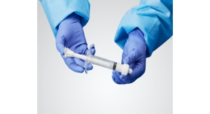
“VIA Disc is intended for use as an allograft to supplement degenerated intervertebral discs,” said Kristine Jacques, vice president and general manager of Interventional Pain Therapies for VIVEX. “Allograft transplantation has long been accepted as an effective option to treat a wide variety of orthopedic and spine-related issues. Age-related wear and tear of an intervertebral disc can cause loss of hydration and degeneration. This process leaves your discs vulnerable to motion, stress, strains your spinal nerves, and can result in lower back pain. VIA Disc is injected into the center of the nucleus in a degenerated disc through a 22-gauge spine needle. After injection, VIA Disc supplements the degenerated disc.”
VIVEX published initial clinical trial (VAST Clinical Trial: Safely Supplementing Tissue Lose to Degenerative Disc Disease) results for VIA Disc in The International Journal of Spine Surgery in April. The paper was the first-ever published 12-month results from Level 1 intradiscal study for treating patients with one or two level symptomatic DDD. Patients with DDD treated with VIA Disc showed improved pain and function greater than 70 percent according to visual analog scale and Oswestry Disability Index, and the improvements were durable one year after the procedure.
“I am highly encouraged by the initial results and safety data from the VAST study. Implantation of disc tissue allograft can be done safely, and early evidence suggests greater than 70 percent improvements in VAS and ODI sustained at 12 months in the allograft groups,” said Douglas Beall, M.D., chief of radiology services at the Clinical Radiology of Oklahoma and principal investigator of the VAST study. “Interventions like VIA Disc have the potential for treating mild-to-moderate degenerative discs that greatly advance an alternative to surgical intervention.”
Orthopedic giant and ODT Top 10 leader Stryker Corporation followed its typical strategy of value-enhancing bolt-on acquisitions, buying Mobius Imaging and sister company Cardan Robotics late last year. Mobius Imaging brings advanced imaging technologies that can be easily integrated into medical workflow, boosting the clinician’s ability to acquire high-quality images. Specifically, Stryker added the Airo TruCT scanner, a mobile, real-time, diagnostic-quality CT imaging system. Prior to being welcomed into Stryker’s fold, Cardan Robotics developed innovative robotics and navigation technology systems for surgical and interventional radiology procedures.
“This acquisition brings expertise in advanced imaging and robotics as well as a robust product pipeline that add to Stryker’s portfolio and will allow the Spine division to provide more complete procedural solutions, including sales, service, and support,” said Spencer Stiles, Stryker’s group president, Orthopaedics and Spine. “We look forward to working together to advance Stryker’s mission to make healthcare better and accelerate our pursuit of category leadership in Neurotechnology, Orthopaedics, and Spine.”
Late last year also saw FDA clearance for the firm’s SAHARA Lateral 3D expandable interbody system, which was added to the franchise as a result of last year’s K2M acquisition. According to Stryker, it is the first ever 3D-printed lateral expandable fusion device. It touts passive expansion capabilities, which means the implant can either be adjusted from a lateral approach intraoperatively or passively adjusted in a staged posterior approach following osteotomy. “Surgeons performing lateral spinal fusion often require versatility to help them achieve optimal outcomes for their patients,” said Gregory Poulter, an orthopedic surgeon at OrthoIndy in Indianapolis. “Stryker’s SAHARA Lateral, with its expansion mechanism that is both actively adjustable from a lateral approach and passively adjustable during a staged posterior procedure, provides an excellent 3D-printed option to help these patients.”
The implant features proprietary Lamellar 3D Titanium Technology, an advanced 3D printing method to build structures previously difficult or impossible to make via traditional manufacturing techniques. It begins with a titanium powder, then SAHARA implants are grown through selective application of a high-energy laser beam to incorporate complex internal geometries and roughened surface architecture. According to the company, roughened titanium surfaces demonstrate increased protein expression compared to smooth surfaces.
NuVasive’s Advanced Materials Science (AMS) portfolio aims to improve spinal fusion by altering the surface and structure of materials with porosity. The segment of the San Diego-based spine technology firm (and longtime ODT Top 10 inhabitant) develops surface and structural technologies to enhance implant material osseointegration and biomechanical properties. The AMS business also designs technologies that improve visualization on a variety of imaging modalities.
The COHERE (cervical spine) and COALESCE (TLIF and PLIF approaches) implants, for example, use porous PEEK technology manufactured through a proprietary extrusion process. Their porous architecture promotes bone in-growth while retaining PEEK’s biomechanical and imaging properties. Modulus titanium interbody implants make up the remainder of NuVasive’s AMS portfolio. Modulus implants combine endplate porosity with optimized body lattice structure for a fully porous architecture and favorable bone in-growth environment.
The Modulus XLIF Dual Sided Plate was launched in May. The low-profile, anti-migration plate enables two fixation points in XLIF surgeries. Surgeons are provided more individualized solutions as a result, especially for multi-stage procedures or in the case of an anterior longitudinal ligament release. The implant’s instrumentation was also simplified so the plate can be placed either before or after the Modulus XLIF implant has been inserted. Modulus ALIF (anterior lumbar interbody fusion) also earned FDA clearance in May. The low-profile implant—the company’s first AMS offering for ALIF—can be used in both lateral and supine positions.
“Since using the Modulus XLIF implant, I have seen extremely favorable clinical results with my patients,” said Mark Wang, an orthopedic spine surgeon at Spine Institute of Arizona. “Now, with the dual sided plate, I can use Modulus XLIF and add a plate when the patient and pathologic features call for it. NuVasive’s portfolio equips me with the appropriate tools to meet the needs of my patients.”
The FDA nod for Cohere TLIF-O rounded out May’s new product trifecta. The porous PEEK implant was engineered for TLIF (transforaminal lumbar interbody fusion). Cohere TLIF-O features NuVasive’s single-step insert and rotate technique and a lordotic design feature in the oblique plane. This permits surgeons to restore spinal sagittal alignment without introducing an undesired coronal misalignment.
The company targeted adolescent idiopathic scoliosis (AIS) with June’s U.S. release of the Reline 3D posterior fixation system. Surgery for AIS involves intricate techniques to address the three-dimensional spinal deformity. The system consolidates multi-step, single-plane correction into one holistic procedure to reduce inefficiency in the OR. 3D adds to the already comprehensive suite of Reline posterior fixation technologies that includes Open, MAS, Trauma, and Small Stature variants.
“Prior to Reline 3D, my approach to treating three-dimensional spinal deformities in pediatrics was inefficient and required treating the coronal, sagittal, and axial planes separately,” said Dr. Robert Cho, chief of staff and pediatric orthopedic surgeon at Shriners for Children Medical Center in Pasadena, Calif. “Reline 3D allows me to simultaneously correct all three planes of the deformity at once, minimizing stress on the bone-screw interface and rod deformation, and maximizing my three-dimensional correction in patients with complex spinal curvatures.”
Every year, there are about 1.62 million instrumented spinal procedures performed in the U.S. alone. Those are usually done using a freehand technique, which can lead to suboptimal outcomes like inaccurate screw positioning, neurological complications due to screw malpositioning, and reoperation to reposition screws. All of these can provoke more surgery related complications.
Traditional navigation systems significantly boost spine surgery outcomes. There is more than a 95 percent success rate as a result of improved accuracy.1 They also reduce screw insertion time by 50 percent2 and decrease X-ray radiation by 92 percent.3 Unfortunately, only 9 percent4 of spine surgeons routinely use them and 66 percent never do. This can be due to a number of factors, including surgeons finding them uncomfortable; they don’t meet surgeons’ time efficiency expectations; ease of use and integration into surgical workflow; and they distract surgeons from patients by requiring them to view a remote screen across the room or at the patient’s feet.
“Spinal navigation and spinal robotics provide the promise of increased accuracy of implant placement,” commented Daniel Sciubba, M.D., professor of neurosurgery at Johns Hopkins University. “However, navigation is associated with surgeon attention drift, which means the surgeon’s attention is diverted away from the patient during the navigated maneuvers. This drift may be unsafe for the patient. In addition, spinal robotics have provided a very cumbersome technique to place hardware safely; namely, robotics require another complex tool between the surgeon and the patient, often at the expense of efficiency and safety.”
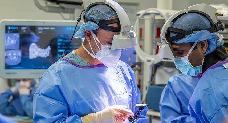
Chicago-based Augmedics has introduced the xvision Spine system, the first augmented reality navigation system to be used in surgery, to provide a solution to these challenges. xvision lets surgeons visualize patients’ 3D anatomy during surgery as if they had “x-ray vision” so they can accurately navigate instruments and implants while looking at the patient, instead of a remote screen.
The system consists of a transparent near-eye display headset and all elements of a traditional navigation system. It accurately determines the position of surgical tool in real-time, then superimposes a virtual trajectory on the patient’s CT data. The 2D and 3D navigation data is projected onto the surgeon’s retina using the headset, allowing them to simultaneously view the patient without averting the eyes to a remote screen. The surgeon is endowed with better control and visualization, leading to potentially easier, faster, and safer surgeries.
“Specifically, the patient will have a registration marker rigidly attached to their spine, and 3D intraoperative imaging will be done,” said Nissan Elimelech, co-founder and CEO of Augmedics. “Augmedics’ software determines where the anatomy is in relation to the registration marker and this information is then transferred wirelessly to the headset. The patient’s anatomy is projected from the transparent augmented reality lenses on the headset to the surgeon’s retina, so they can see the data while looking at the patient. Its wearable guidance technology reduces traditional line of sight issues between the IR camera and markers and provides a linear working environment within the surgeon’s field of sight with the AR headset.”
“The platform basically includes the headset and a patient reference frame, the latter of which is a small spine clamp,” added Dr. Sciubba. “It does not include a large robot or a navigation system with multiple cameras and monitors that are routinely blocked or offline during the operation. In this way, the surgeon can move quickly through surgery without the extra workflow friction of robotics and traditional navigation. Nonetheless, the surgeon can use navigation whenever desired. A wireless foot pedal can turn the AR on and off whenever needed. So, the surgeon can move quickly and safely, harnessing the benefits of navigation without the usual workflow inefficiencies.”
The xvision Spine system achieved FDA 510(k) clearance and was launched in the U.S. in December. In June 2020, Dr. Sciubba and a team of Johns Hopkins University surgeons became the first to use xvision in a successful procedure—a spinal fusion surgery.
“The system has been very intuitive to use,” explained Dr. Sciubba. “Our first patient was an open lumbar decompression and fusion, which went flawlessly. That very same week, we used the xvision during an en bloc resection of a spinal chordoma, often considered one of the most challenges surgeries in spine care. We saw no challenges in either case within the same week. We expect both novice and expert surgeons to easily adopt this intuitive system.”
Augmedics plans to earn a CE mark by the end of next year, and aims to commercialize the xvision Spine system worldwide by the end of 2022. Elimelech said that Augmedics is continuing to explore additional indications for xvision, including cranial surgery, joint reconstruction, and knee and hip replacements.
“The xvision is currently being used by 10 surgeons in four facilities,” said Elimelech. “To date we have heard nothing but excellent responses from the surgeons who have used the system. The feedback includes things like “amazing” and “game-changing technology,” as well as “very intuitive.” When it is used for minimally-invasive procedures, surgeons understand that the system is so valuable because they are looking right at the patient rather than at a remote screen, and they can see everything through the skin as if it were an open procedure.”
Tools of the Trade
Surgical instrumentation for spine procedures is increasingly driven by more complexity, multifunctionality, ease of use, and human factors engineering. The market continues to expand for both new and innovative instruments to improve the end-user experience, both for surgeons and patients. Spine OEMs and contract instrument manufacturers work closely with surgeons on instrument designs to improve ease of use and ergonomics.
“Our products are designed with the deliberate intent of customization in mind from the start,” explained Chad Ryshkus, director of marketing and product development at MedTorque, a Kenosha, Wis.-based manufacturer of orthopedic instruments and implants, including single-procedure torque-limiting drivers. “Although we have a broad offering of standard spine instrument designs to choose from, we know through experience that our OEM customers often need to tailor these instruments to meet their customer needs. Our design team works closely with our customer partners to ensure those needs are met.”
Development efforts of most OEMs have typically been focused on the implant and procedure, and less so on the instrumentation. However, today’s technology has prompted innovation to instrumentation in the field of spine surgery. Of the many instruments used during a spinal procedure, torque-limiting instruments result from applying a simple technology to a specific need.
Torque-limiting instruments use a mechanism that limits torque applied during the tightening of fasteners like implant screws. The mechanism is typically housed within the instrument’s handle, and is designed to slip after the predetermined torque load has been reached. This slip action is comparable to that of other clutch mechanisms like impact wrenches and electric screwdrivers.
“Torque-limiting products designed for use in orthopedic and spine surgery are most often used to limit the amount of torque that is applied to a fastener that will be implanted in the body,” said Ryshkus. “In spinal surgery, for example, the need to control the amount of applied torque is critical to the function of the implant. Rods, such as those used in posterior lumbar fusion, are most often held in place by set screws tightened to a specific torque value. If the set screw is not tightened enough, the implant may loosen and cause harm to the patient. If the set screw is tightened with too much force (torque) it could damage the implant.
Incorporating a positive limit on applied torque reduces a large amount of the surgeon’s guesswork. Due to its effectiveness, controlling torque load is a straightforward approach for most applications. Engineers can specify the tightening torque of threaded fasteners instead of the actual clamping force generated by the screw, which can be tough to measure in the OR. By relying on this method, however, torque-limiting instrumentation must be incredibly reliable. Performance anywhere outside the narrow range of acceptable variability may compromise the outcome of the surgery.
“We understand our customers and innovate with experience to create high quality, precision instruments for the orthopedic and spine industry,” said Ryshkus. “Our design engineers have decades of experience in designing and co-developing highly engineered, complex instrument assemblies for spine and orthopedic surgery.”
References
The newly born SeaSpine charted its course toward interbody devices, minimally invasive surgery technologies, and the IsoTis orthobiologics brand—a range of osteoconductive and osteoinductive demineralized bone and synthetic matrix products. A favorable wind filled the firm’s sails as well—HealthPointCapital anticipated $140 million in first year revenue and the company touted a strong balance sheet with $47 million of cash and no debt post spinoff.
SeaSpine’s precious cargo is its proprietary NanoMetalene surface technology. Using high-energy, low-temperature atomic fusion deposition, a sub-micron layer of commercially pure titanium is molecularly bonded to a polyetheretherketone (PEEK) interbody implant. The result is a bone-friendly titanium endplate surface throughout the apertures with the traditional PEEK upshots: biocompatibility, a modulus of elasticity that mimics bone, and radiographic visibility for post-op imaging. Last July, SeaSpine celebrated the 20,000th implantation of a NanoMetalene interbody implant.
“This milestone reflects the success of our strategy to align NanoMetalene with our DBM portfolio and deliver synergistic solutions that maximize clinical value while reducing the need for high-cost orthobiologics,” Troy Woolley, vice president of marketing, Spinal Implants for SeaSpine, told the press.
Many of SeaSpine’s fleet of spine implants reflect its nautical moniker: Regatta (lateral lumbar interbody fusion), Shoreline (anterior cervical fixation/fusion), Mariner (posterior thoracolumbar fixation), Pacifica (posterior/transforaminal lumbar interbody fusion), Malibu (pedicle screws for posterior lumbar fusion), and Atoll (OCT spinal system for posterior cervical fusion).
The company’s fleet continues to grow throughout this year despite the pandemic’s ill winds blowing spine companies way off course. 2020 has seen seven products set sail into the spine market so far: the Mariner Midline posterior fixation system in January, the NorthStar OCT and cervical facet fusion systems after a COVID-19 hiatus in June, the Mariner MIS and Outrigger spinal fixation systems in July, and both the Explorer TO expandable interbody and Shoreline RT (Reef Topography) cervical interbody implant system. Reef Topography is a new technique that applies machined macrostructures and undercut features to promote bony interlocking. The process increases NanoMetalene endplate surface area in the endplate by 40 percent, and in the apertures by 50 percent, according to the company.
Changing course, VIVEX Biologics is an Atlanta, Ga.-based regenerative medicine company that specializes in development of naturally sourced treatment options, and it created the VIA Disc to provide a viable treatment option. VIA Disc is a non-surgical, injectable treatment option for patients suffering from chronic lower back pain resulting from DDD.
Lower back pain is one of the most expensive occupational disorders in the U.S. and the leading cause of disability globally. Degenerative disc disease (DDD) is a major factor contributing to this disability, and is the most common source of low back pain in adults. Today there are limited treatment options for DDD, including progression to opioids and surgery for patients who fail more conservative management.
“VIA Disc is intended for use as an allograft to supplement degenerated intervertebral discs,” said Kristine Jacques, vice president and general manager of Interventional Pain Therapies for VIVEX. “Allograft transplantation has long been accepted as an effective option to treat a wide variety of orthopedic and spine-related issues. Age-related wear and tear of an intervertebral disc can cause loss of hydration and degeneration. This process leaves your discs vulnerable to motion, stress, strains your spinal nerves, and can result in lower back pain. VIA Disc is injected into the center of the nucleus in a degenerated disc through a 22-gauge spine needle. After injection, VIA Disc supplements the degenerated disc.”
VIVEX published initial clinical trial (VAST Clinical Trial: Safely Supplementing Tissue Lose to Degenerative Disc Disease) results for VIA Disc in The International Journal of Spine Surgery in April. The paper was the first-ever published 12-month results from Level 1 intradiscal study for treating patients with one or two level symptomatic DDD. Patients with DDD treated with VIA Disc showed improved pain and function greater than 70 percent according to visual analog scale and Oswestry Disability Index, and the improvements were durable one year after the procedure.
“I am highly encouraged by the initial results and safety data from the VAST study. Implantation of disc tissue allograft can be done safely, and early evidence suggests greater than 70 percent improvements in VAS and ODI sustained at 12 months in the allograft groups,” said Douglas Beall, M.D., chief of radiology services at the Clinical Radiology of Oklahoma and principal investigator of the VAST study. “Interventions like VIA Disc have the potential for treating mild-to-moderate degenerative discs that greatly advance an alternative to surgical intervention.”
Orthopedic giant and ODT Top 10 leader Stryker Corporation followed its typical strategy of value-enhancing bolt-on acquisitions, buying Mobius Imaging and sister company Cardan Robotics late last year. Mobius Imaging brings advanced imaging technologies that can be easily integrated into medical workflow, boosting the clinician’s ability to acquire high-quality images. Specifically, Stryker added the Airo TruCT scanner, a mobile, real-time, diagnostic-quality CT imaging system. Prior to being welcomed into Stryker’s fold, Cardan Robotics developed innovative robotics and navigation technology systems for surgical and interventional radiology procedures.
“This acquisition brings expertise in advanced imaging and robotics as well as a robust product pipeline that add to Stryker’s portfolio and will allow the Spine division to provide more complete procedural solutions, including sales, service, and support,” said Spencer Stiles, Stryker’s group president, Orthopaedics and Spine. “We look forward to working together to advance Stryker’s mission to make healthcare better and accelerate our pursuit of category leadership in Neurotechnology, Orthopaedics, and Spine.”
Late last year also saw FDA clearance for the firm’s SAHARA Lateral 3D expandable interbody system, which was added to the franchise as a result of last year’s K2M acquisition. According to Stryker, it is the first ever 3D-printed lateral expandable fusion device. It touts passive expansion capabilities, which means the implant can either be adjusted from a lateral approach intraoperatively or passively adjusted in a staged posterior approach following osteotomy. “Surgeons performing lateral spinal fusion often require versatility to help them achieve optimal outcomes for their patients,” said Gregory Poulter, an orthopedic surgeon at OrthoIndy in Indianapolis. “Stryker’s SAHARA Lateral, with its expansion mechanism that is both actively adjustable from a lateral approach and passively adjustable during a staged posterior procedure, provides an excellent 3D-printed option to help these patients.”
The implant features proprietary Lamellar 3D Titanium Technology, an advanced 3D printing method to build structures previously difficult or impossible to make via traditional manufacturing techniques. It begins with a titanium powder, then SAHARA implants are grown through selective application of a high-energy laser beam to incorporate complex internal geometries and roughened surface architecture. According to the company, roughened titanium surfaces demonstrate increased protein expression compared to smooth surfaces.
NuVasive’s Advanced Materials Science (AMS) portfolio aims to improve spinal fusion by altering the surface and structure of materials with porosity. The segment of the San Diego-based spine technology firm (and longtime ODT Top 10 inhabitant) develops surface and structural technologies to enhance implant material osseointegration and biomechanical properties. The AMS business also designs technologies that improve visualization on a variety of imaging modalities.
The COHERE (cervical spine) and COALESCE (TLIF and PLIF approaches) implants, for example, use porous PEEK technology manufactured through a proprietary extrusion process. Their porous architecture promotes bone in-growth while retaining PEEK’s biomechanical and imaging properties. Modulus titanium interbody implants make up the remainder of NuVasive’s AMS portfolio. Modulus implants combine endplate porosity with optimized body lattice structure for a fully porous architecture and favorable bone in-growth environment.
The Modulus XLIF Dual Sided Plate was launched in May. The low-profile, anti-migration plate enables two fixation points in XLIF surgeries. Surgeons are provided more individualized solutions as a result, especially for multi-stage procedures or in the case of an anterior longitudinal ligament release. The implant’s instrumentation was also simplified so the plate can be placed either before or after the Modulus XLIF implant has been inserted. Modulus ALIF (anterior lumbar interbody fusion) also earned FDA clearance in May. The low-profile implant—the company’s first AMS offering for ALIF—can be used in both lateral and supine positions.
“Since using the Modulus XLIF implant, I have seen extremely favorable clinical results with my patients,” said Mark Wang, an orthopedic spine surgeon at Spine Institute of Arizona. “Now, with the dual sided plate, I can use Modulus XLIF and add a plate when the patient and pathologic features call for it. NuVasive’s portfolio equips me with the appropriate tools to meet the needs of my patients.”
The FDA nod for Cohere TLIF-O rounded out May’s new product trifecta. The porous PEEK implant was engineered for TLIF (transforaminal lumbar interbody fusion). Cohere TLIF-O features NuVasive’s single-step insert and rotate technique and a lordotic design feature in the oblique plane. This permits surgeons to restore spinal sagittal alignment without introducing an undesired coronal misalignment.
The company targeted adolescent idiopathic scoliosis (AIS) with June’s U.S. release of the Reline 3D posterior fixation system. Surgery for AIS involves intricate techniques to address the three-dimensional spinal deformity. The system consolidates multi-step, single-plane correction into one holistic procedure to reduce inefficiency in the OR. 3D adds to the already comprehensive suite of Reline posterior fixation technologies that includes Open, MAS, Trauma, and Small Stature variants.
“Prior to Reline 3D, my approach to treating three-dimensional spinal deformities in pediatrics was inefficient and required treating the coronal, sagittal, and axial planes separately,” said Dr. Robert Cho, chief of staff and pediatric orthopedic surgeon at Shriners for Children Medical Center in Pasadena, Calif. “Reline 3D allows me to simultaneously correct all three planes of the deformity at once, minimizing stress on the bone-screw interface and rod deformation, and maximizing my three-dimensional correction in patients with complex spinal curvatures.”
Every year, there are about 1.62 million instrumented spinal procedures performed in the U.S. alone. Those are usually done using a freehand technique, which can lead to suboptimal outcomes like inaccurate screw positioning, neurological complications due to screw malpositioning, and reoperation to reposition screws. All of these can provoke more surgery related complications.
Traditional navigation systems significantly boost spine surgery outcomes. There is more than a 95 percent success rate as a result of improved accuracy.1 They also reduce screw insertion time by 50 percent2 and decrease X-ray radiation by 92 percent.3 Unfortunately, only 9 percent4 of spine surgeons routinely use them and 66 percent never do. This can be due to a number of factors, including surgeons finding them uncomfortable; they don’t meet surgeons’ time efficiency expectations; ease of use and integration into surgical workflow; and they distract surgeons from patients by requiring them to view a remote screen across the room or at the patient’s feet.
“Spinal navigation and spinal robotics provide the promise of increased accuracy of implant placement,” commented Daniel Sciubba, M.D., professor of neurosurgery at Johns Hopkins University. “However, navigation is associated with surgeon attention drift, which means the surgeon’s attention is diverted away from the patient during the navigated maneuvers. This drift may be unsafe for the patient. In addition, spinal robotics have provided a very cumbersome technique to place hardware safely; namely, robotics require another complex tool between the surgeon and the patient, often at the expense of efficiency and safety.”
Chicago-based Augmedics has introduced the xvision Spine system, the first augmented reality navigation system to be used in surgery, to provide a solution to these challenges. xvision lets surgeons visualize patients’ 3D anatomy during surgery as if they had “x-ray vision” so they can accurately navigate instruments and implants while looking at the patient, instead of a remote screen.
The system consists of a transparent near-eye display headset and all elements of a traditional navigation system. It accurately determines the position of surgical tool in real-time, then superimposes a virtual trajectory on the patient’s CT data. The 2D and 3D navigation data is projected onto the surgeon’s retina using the headset, allowing them to simultaneously view the patient without averting the eyes to a remote screen. The surgeon is endowed with better control and visualization, leading to potentially easier, faster, and safer surgeries.
“Specifically, the patient will have a registration marker rigidly attached to their spine, and 3D intraoperative imaging will be done,” said Nissan Elimelech, co-founder and CEO of Augmedics. “Augmedics’ software determines where the anatomy is in relation to the registration marker and this information is then transferred wirelessly to the headset. The patient’s anatomy is projected from the transparent augmented reality lenses on the headset to the surgeon’s retina, so they can see the data while looking at the patient. Its wearable guidance technology reduces traditional line of sight issues between the IR camera and markers and provides a linear working environment within the surgeon’s field of sight with the AR headset.”
“The platform basically includes the headset and a patient reference frame, the latter of which is a small spine clamp,” added Dr. Sciubba. “It does not include a large robot or a navigation system with multiple cameras and monitors that are routinely blocked or offline during the operation. In this way, the surgeon can move quickly through surgery without the extra workflow friction of robotics and traditional navigation. Nonetheless, the surgeon can use navigation whenever desired. A wireless foot pedal can turn the AR on and off whenever needed. So, the surgeon can move quickly and safely, harnessing the benefits of navigation without the usual workflow inefficiencies.”
The xvision Spine system achieved FDA 510(k) clearance and was launched in the U.S. in December. In June 2020, Dr. Sciubba and a team of Johns Hopkins University surgeons became the first to use xvision in a successful procedure—a spinal fusion surgery.
“The system has been very intuitive to use,” explained Dr. Sciubba. “Our first patient was an open lumbar decompression and fusion, which went flawlessly. That very same week, we used the xvision during an en bloc resection of a spinal chordoma, often considered one of the most challenges surgeries in spine care. We saw no challenges in either case within the same week. We expect both novice and expert surgeons to easily adopt this intuitive system.”
Augmedics plans to earn a CE mark by the end of next year, and aims to commercialize the xvision Spine system worldwide by the end of 2022. Elimelech said that Augmedics is continuing to explore additional indications for xvision, including cranial surgery, joint reconstruction, and knee and hip replacements.
“The xvision is currently being used by 10 surgeons in four facilities,” said Elimelech. “To date we have heard nothing but excellent responses from the surgeons who have used the system. The feedback includes things like “amazing” and “game-changing technology,” as well as “very intuitive.” When it is used for minimally-invasive procedures, surgeons understand that the system is so valuable because they are looking right at the patient rather than at a remote screen, and they can see everything through the skin as if it were an open procedure.”
Tools of the Trade
Surgical instrumentation for spine procedures is increasingly driven by more complexity, multifunctionality, ease of use, and human factors engineering. The market continues to expand for both new and innovative instruments to improve the end-user experience, both for surgeons and patients. Spine OEMs and contract instrument manufacturers work closely with surgeons on instrument designs to improve ease of use and ergonomics.
“Our products are designed with the deliberate intent of customization in mind from the start,” explained Chad Ryshkus, director of marketing and product development at MedTorque, a Kenosha, Wis.-based manufacturer of orthopedic instruments and implants, including single-procedure torque-limiting drivers. “Although we have a broad offering of standard spine instrument designs to choose from, we know through experience that our OEM customers often need to tailor these instruments to meet their customer needs. Our design team works closely with our customer partners to ensure those needs are met.”
Development efforts of most OEMs have typically been focused on the implant and procedure, and less so on the instrumentation. However, today’s technology has prompted innovation to instrumentation in the field of spine surgery. Of the many instruments used during a spinal procedure, torque-limiting instruments result from applying a simple technology to a specific need.
Torque-limiting instruments use a mechanism that limits torque applied during the tightening of fasteners like implant screws. The mechanism is typically housed within the instrument’s handle, and is designed to slip after the predetermined torque load has been reached. This slip action is comparable to that of other clutch mechanisms like impact wrenches and electric screwdrivers.
“Torque-limiting products designed for use in orthopedic and spine surgery are most often used to limit the amount of torque that is applied to a fastener that will be implanted in the body,” said Ryshkus. “In spinal surgery, for example, the need to control the amount of applied torque is critical to the function of the implant. Rods, such as those used in posterior lumbar fusion, are most often held in place by set screws tightened to a specific torque value. If the set screw is not tightened enough, the implant may loosen and cause harm to the patient. If the set screw is tightened with too much force (torque) it could damage the implant.
Incorporating a positive limit on applied torque reduces a large amount of the surgeon’s guesswork. Due to its effectiveness, controlling torque load is a straightforward approach for most applications. Engineers can specify the tightening torque of threaded fasteners instead of the actual clamping force generated by the screw, which can be tough to measure in the OR. By relying on this method, however, torque-limiting instrumentation must be incredibly reliable. Performance anywhere outside the narrow range of acceptable variability may compromise the outcome of the surgery.
“We understand our customers and innovate with experience to create high quality, precision instruments for the orthopedic and spine industry,” said Ryshkus. “Our design engineers have decades of experience in designing and co-developing highly engineered, complex instrument assemblies for spine and orthopedic surgery.”
References